
 685 738 325

Pet ultrasound and X-ray
Diagnostic imaging at our clinic in Gràcia, Barcelona: same-visit results for dogs and cats.
What it is
Looking inside without opening up
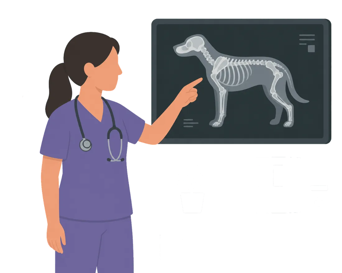
Veterinary diagnostic imaging allows evaluation of internal structures — bones, organs, tissue — without exploratory surgery. It is an essential diagnostic tool that complements the physical examination and blood tests, and in many cases provides the definitive diagnosis.
At Rovira Veterinary Clinic, in Gràcia, Barcelona, we have digital X-ray and ultrasound on site. Results are available at the same visit, allowing immediate clinical decisions without unnecessary delays.
The two techniques
X-ray and ultrasound: different tools for different problems

Digital X-ray
The technique of choice for the musculoskeletal system, thorax and detection of foreign bodies. Provides high-resolution images immediately.
- Fractures and luxations
- Pulmonary and cardiac pathology
- Gastrointestinal obstructions
- Bone tumours
- Post-operative monitoring

Ultrasound
No radiation, in real time. Allows evaluation of abdominal organs and guides aspirates or biopsies with precision. An indispensable technique in soft-tissue medicine.
- Liver, kidneys, spleen and bladder
- Reproductive system
- Abdominal nodules and masses
- Guided aspirates and biopsies
- Pregnancy monitoring
When we use it
Typical situations where we recommend imaging

Emergency or trauma
Accidents & trauma
Road accidents, falls or fights with suspected fracture, internal haemorrhage or other injuries requiring immediate assessment.

Internal symptoms
Abdominal or thoracic pain
Vomiting, bloating, breathing difficulty or pain on palpation are common indications for requesting imaging.

Progress monitoring
Follow-up of chronic conditions
Monitoring of tumours, renal disease, heart conditions or any process requiring periodic radiological review.
How we work
From scan to diagnosis in a single visit
One of the advantages of on-site diagnostic imaging is immediacy. We don't make you wait days for results — we interpret images on the spot and explain the findings before you leave the consultation.
In Gràcia, Barcelona, diagnostic imaging is part of the standard consultation flow — it is not a separate service requiring a referral or a specific appointment at another centre.
- 1 Prior clinical assessment to determine which test is appropriate
- 2 Positioning of the animal with minimum possible stress
- 3 High-resolution digital images obtained
- 4 Immediate interpretation by the vet in consultation
- 5 Findings explained to the owner with the images
- 6 Report and action plan based on the results
Frequently asked questions
What people ask us most about diagnostic imaging
Is X-ray dangerous for my pet?
Veterinary X-rays use very low radiation doses, equivalent to those of a transatlantic flight. For one-off studies there is no risk. In patients requiring many X-rays over time we assess exposure on a case-by-case basis to minimise cumulative dose.
Does my pet need sedation for an ultrasound or X-ray?
In most cases it is not necessary. We use low-stress handling techniques that allow us to obtain good images without sedating the animal. In very anxious patients or studies requiring precise positioning we may consider sedation, but always as a last resort.
When do you use X-ray and when ultrasound?
It depends on what we need to see. X-ray is ideal for bones, the thorax and detecting gas or foreign bodies. Ultrasound is better for abdominal soft organs, masses and guiding biopsies. In many cases we use both techniques together to get a complete picture of the patient.
Are results available the same day?
Yes. Both digital X-ray and ultrasound provide immediate results in the consultation. We explain the findings on the spot and give you the images. If the case requires a specialist second opinion, we send it to an external veterinary radiologist and results arrive within 24–48 hours.
Does your pet need an X-ray or ultrasound?
Book an appointment